Fig 3 -.
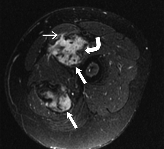
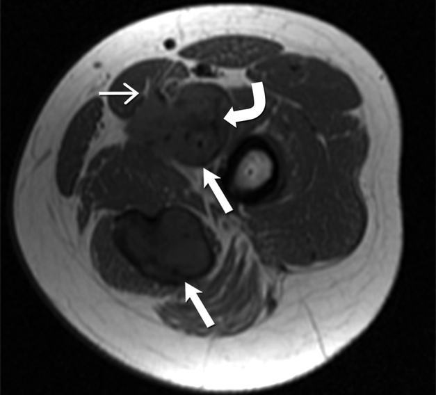
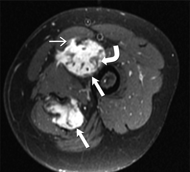
24-year-old-man with desmoid fibromatosis.
(A) Axial T2 weighted MR of left thigh shows two heterogeneous high signal intensity soft tissue masses (straight open arrows) in the intermuscular compartment in the left thigh. Bands of linear low T2 signal intensity are seen within the tumor (curved arrow), reflecting the presence of collagen and low cellularity. Linear extensions are also seen extending from the periphery of the mass along the fascia (straight closed arrow).
(B)Axial T1 weighted MR of the left thigh shows two heterogeneous soft tissue masses (straight open arrows) in the intermuscular compartment in the left thigh, which are isointense to the skeletal muscle. Bands of linear low T1 signal intensity are seen within the tumor (curved arrow), reflecting the presence of collagen and low cellularity. Linear extensions are also seen extending from the periphery of the mass along the fascia (straight closed arrow).
(C) Axial post contrast T1 weighted MR of the left thigh shows two heterogeneous enhancing soft tissue masses (straight open arrows) in the intermuscular compartment in the left thigh. Avid enhancement within the mass reflects areas of high cellularity within the tumor. However, non-enhancing linear bands of low signal intensity are seen within the tumor (curved arrow), reflecting the presence of collagen bands. Linear extensions are also seen extending from the periphery of the mass along the fascia (straight closed arrow).


