Fig. 6-.
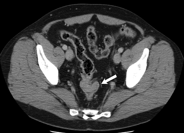
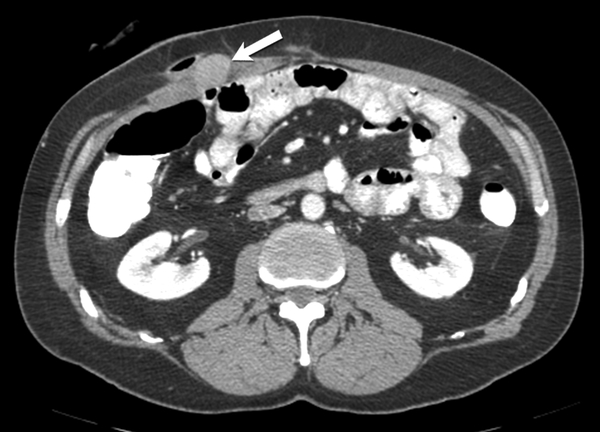
47-year-old man with familial adenomatosis polyposis
(A) Axial contrast enhanced CT of the abdomen shows enhancing circumferential rectal tumor (arrow in A). Biopsy confirmed rectal adenocarcinoma. Patient underwent low anterior resection and diverting ileostomy.
(B) Axial CT scan of the abdomen performed 4 months after surgery shows a 2 cm soft tissue mass in the right abdominal wall near the ileostomy surgical scar. Biopsy of the abdominal wall mass confirmed the diagnosis of desmoid fibromatosis.


