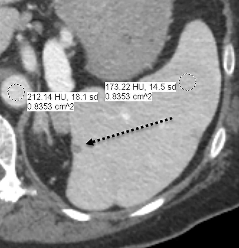
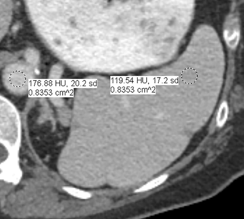
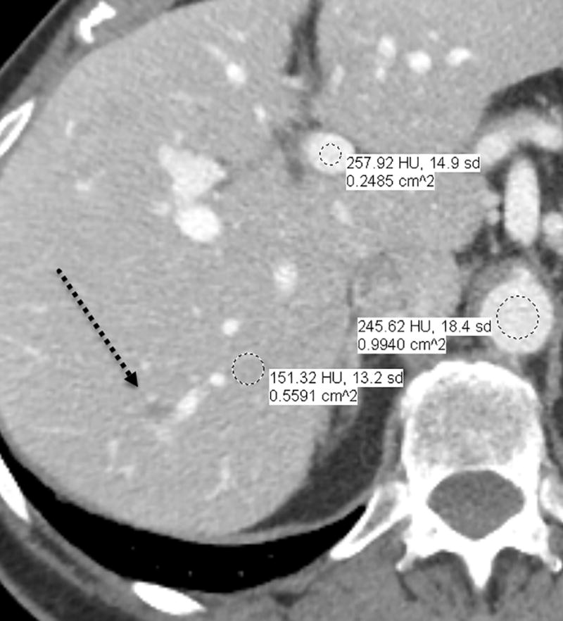
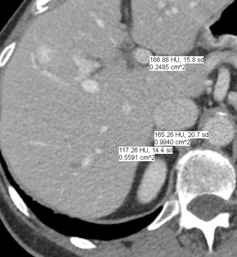
Fig. 1.


a–d—Two case examples from different patients, each demonstrating a small benign finding (a lesion that is unchanged between examinations) for comparison between fixed-dose (a,c) and weight-based dosing (WBD) (b,d) examinations when IV contrast load was allowed below 110 mL (38.5 g of iodine) on the WBD examination. Inferior overall enhancement and lesion depiction was noted qualitatively by readers and upon quantitative measurements for the WBD cases. Readers noticed that a subtle splenic lesion (a, arrow) and the nodular enhancement of a hemangioma (c, arrow) were only barely seen on the WBD examinations.
