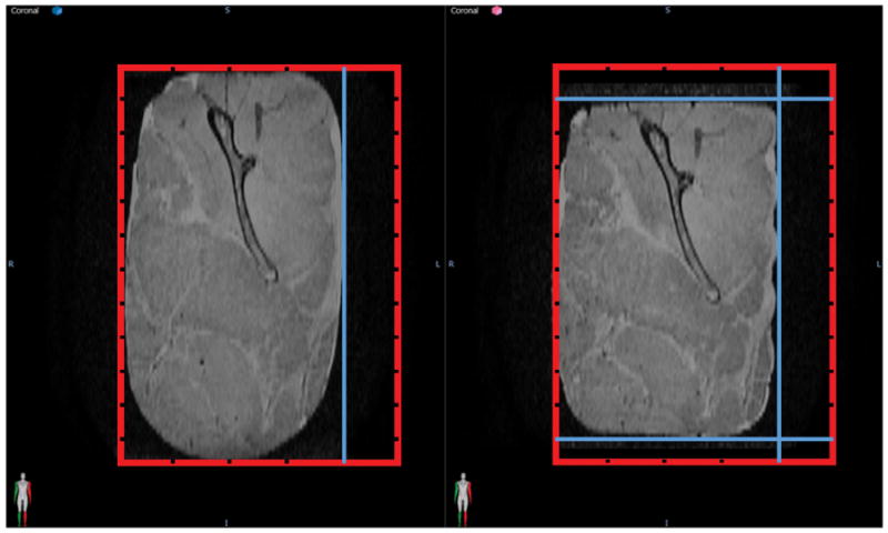
Figure 1. Porcine Phantom Representative Deformation.

A deformation was applied to the porcine phantom by moving the dividers. The red box represents the container with the grooves for the movable dividers and the position of the dividers is shown in blue. The original position is shown on the left and a deformed position using more movable dividers to secure the phantom in place is shown on the right. (Reproduced with permission from Wiley 24.)
