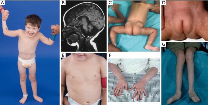
Figure 3.
Clinical photographs of patients with PMM2-CDG. (A) Happy demeanor. Truncal hypotonia and kyphoscoliosis are often present; (B) MRI showing cerebellar vermis hypoplasia; (C) posterior suprapelvic fat pads present in approximately 25% of patients with PMM2-CDG; (D) suprapubic fat pad seen in infancy, usually disappears by 5 years of age; (E) inverted nipples; (F,G) long thin fingers and atrophied low extremities secondary to demyelinating peripheral neuropathy. Courtesy of NIH, Lynne Wolfe, CRNP, Donna Krasnewich, MD, PhD. MRI, magnetic resonance imaging.

