Fig. 6.
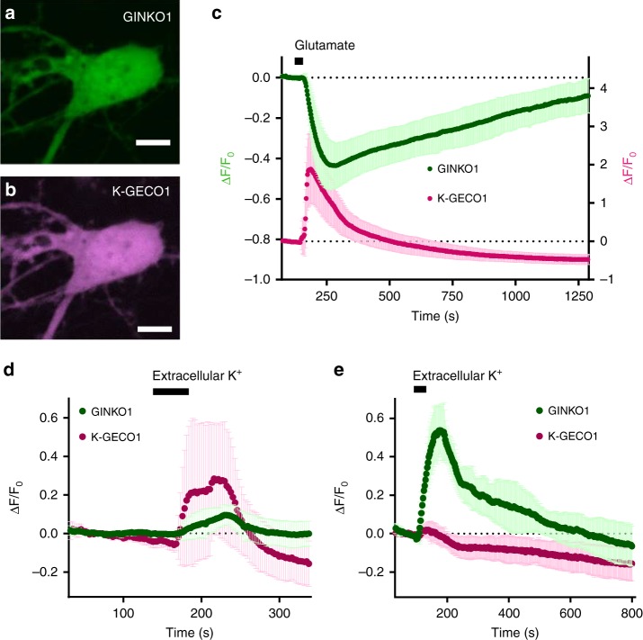
Dual-Color imaging of K+ and Ca2+ dynamics in cultured cortical dissociated neurons and glial cells. a Representative GFP channel fluorescence image of neuron soma region expressing GINKO1 (scale bar = 5 µm). b Representative RFP channel fluorescence image of neuron soma region expressing K-GECO1 (scale bar = 5 µm). c Normalized fluorescence intensity change (ΔF/F0) time course of GINKO1 and K-GECO1 with stimulation (30 s, 500 µM) of glutamate on neurons (n = 10), data are expressed as mean ± SD. d Normalized fluorescence intensity change (ΔF/F0) time course of GINKO1 and K-GECO1 with stimulation by a high concentration (30 mM) of extracellular K+ on neurons (n = 7), data are expressed as mean ± SD. e Normalized fluorescence intensity change (ΔF/F0) time course of GINKO1 and K-GECO1 with stimulation by a high concentration (30 mM) of extracellular K+ on glial cells (n = 4), data are expressed as mean ± SD

