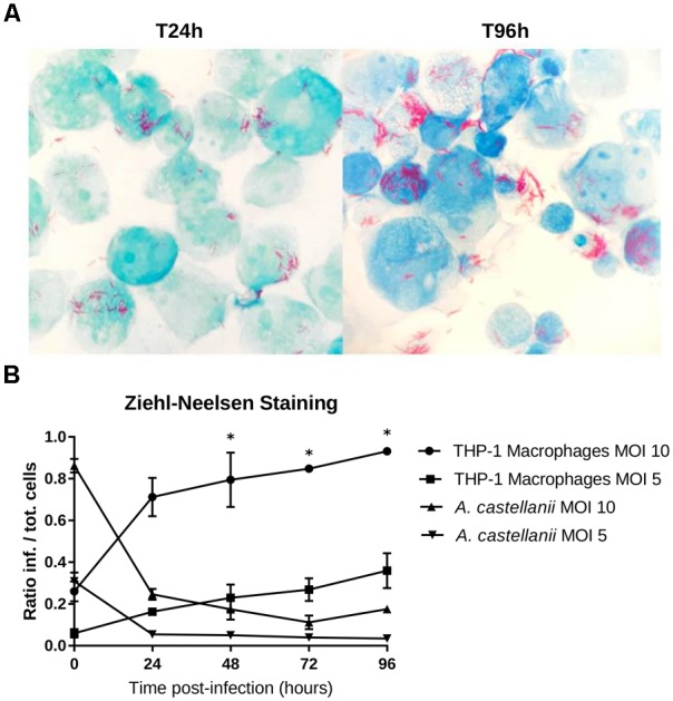
FIGURE 3.

THP-1 macrophage infection shown using Ziehl-Neelsen staining. (A) THP-1 cells strained with ZN 24 and 96 h post-infection with an MOI of 10. Mycobacteria were observed in the extracellular medium, probably explained by recent host cell lysis events; extracellular growth is unlikely to have occurred because infected cells were treated with gentamycin to remove bacteria that were not internalized. (B) Ratio of the infected cells to the total number of cells counted (means ± 2 standard deviations). The asterisks (∗) indicate the timepoints at which each condition was significantly different from another by doing pair-wise chi-squared tests between pairs of triplicates with five degrees of freedom (p-value < 0.05).
