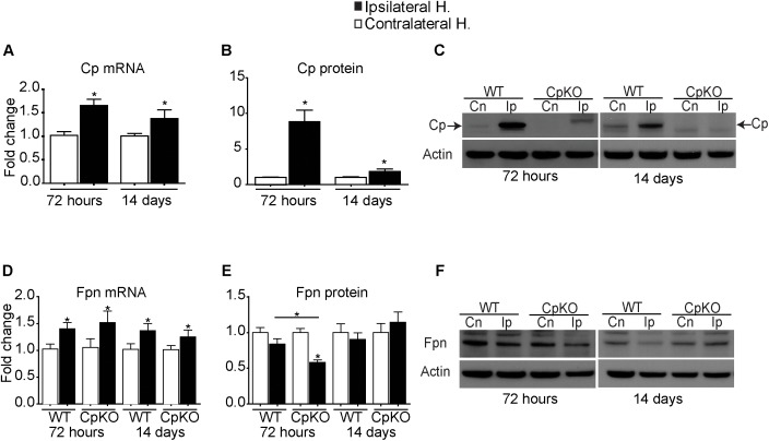
FIGURE 2.
Changes in mRNA detected by q-PCR and protein expression detected by Western blot analysis of Cp (A–C) and Fpn (D–F) in the ipsilateral ischemic side as compared to the contralateral uninjured side at 72 h and 14 days after pMCAO. Changes in the ipsilateral ischemic side are represented as fold-change as compared to contralateral uninjured side. Changes in expression of Cp mRNA (A), and protein (B) quantified from Western blots; a representative example of the Western blot for Cp is shown (C). Note the marked increase in Cp protein at 72 h (8.8-fold) and which remains significantly elevated (twofold) at 14 days. (C) The weak bands seen on the ipsilateral side in the Cp null mice are non-specific bands that are not at the expected 135 kDa GPI-Cp band (arrows). Changes in Fpn mRNA (D) and protein (E) and representative Western blots (F). Fpn is significantly reduced on the ipsilateral side in Cp null mice at 72 h. β actin was used as a loading control. n = 6 per group; ∗p-value ≤ 0.05.

