FIGURE 3.
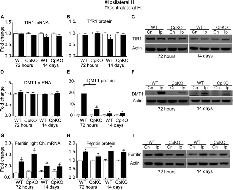
Changes in mRNA and protein expression of TfR1 (A–C), DMT1 (D–F), and ferritin (G–I) shown as fold-change in the ipsilateral ischemic side as compared to the contralateral uninjured side at 72 h and 14 days after pMCAO. No changes are seen in TfR1 mRNA (A) or protein (B) except for a small but significant reduction in the ipsilateral side in wildtype mice at 14 days. Representative Western blot shown in C. Changes in expression of DMT1 mRNA (D), and protein (E) quantified from Western blots; representative Western blot shown in F. Note the marked increase in DMT1 protein on the ipsilateral side in wildtype (19.5-fold) and Cp null (sixfold) mice at 72 h, which remain elevated at twofold at 14 days in both genotypes. Changes in mRNA expression of ferritin light chain (G) and ferritin protein (H) and a representative Western blot (I). Note the increase in mRNA and protein levels in the ipsilateral side in all groups. β actin was used as a loading control; note that the same blot was used to probe DMT1 (F) and Cp (Figure 2C) hence the actin blots for these are the same. n = 6 per group; ∗p-value ≤ 0.05.

