FIGURE 6.
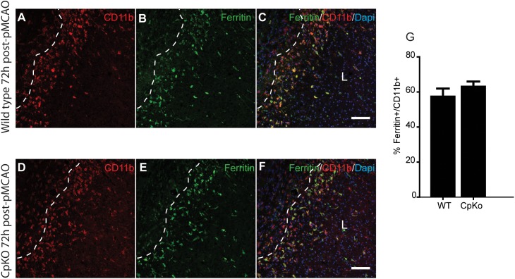
Immunofluorescence labeling for ferritin in macrophage/microglia. Double immunofluorescence labeling show that ferritin+/CD11b+ macrophage/microglia are located mainly in the lesion (L) along the lesion border in both wildtype (A–C) and CpKO (D–F) mice 72 h after pMCAO. Dotted line demarcates the lesion boundary. The merged images (C,F) show CD11b, ferritin and DAPI nuclear staining. Note that the number of CD11b+/ferritin+ doubled labeled cells are not significantly different in the two genotypes (G). Scale bar = 100 μm.

