FIGURE 7.
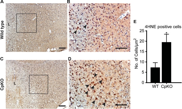
Changes in lipid peroxidation detected by 4-HNE labeling. Tissue sections of wildtype (A,B) and CpKO (C,D) mice showing immunohistochemical staining for 4HNE. The area demarcated by the black squares in A and C are shown at higher magnification in panels B,D. The lesion area in A,C are indicated by “L.” The 4HNE staining is seen as a brown reaction product (arrowheads); the tissue sections were counterstained with Mayer’s hemalaum which labels the cell nuclei blue. (E) Graph shows that the number of 4HNE+ cells is significantly higher in CpKO as compared to wildtype mice. Scale bar = 100 μm, ∗p-value ≤ 0.05.

