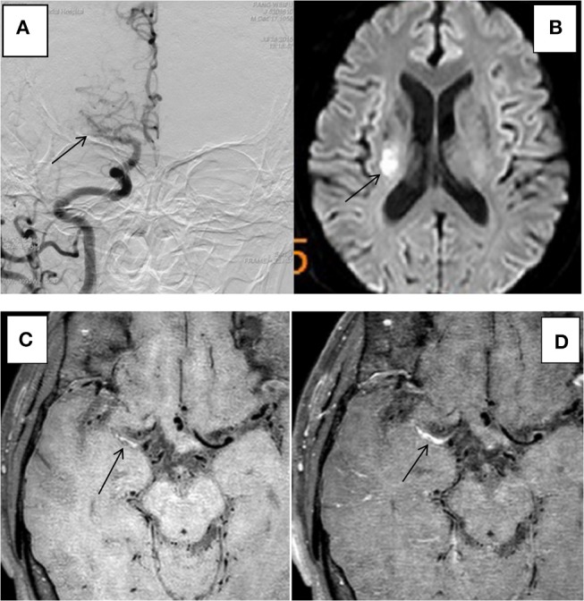
Figure 3.

HR-MRI of a culprit MCA occlusion in an 59-years-oldmale. (A) DSA showed occlusion of the right MCA. (B) DWI showed that the right half of the oval center was limited by diffusion. (C) HR-MRI Tl WI showed plaque formation in the Ml segment of MCA. (D) High-resolution enhanced Tl WI showed marked enhancement of the Ml segment of MCA.
