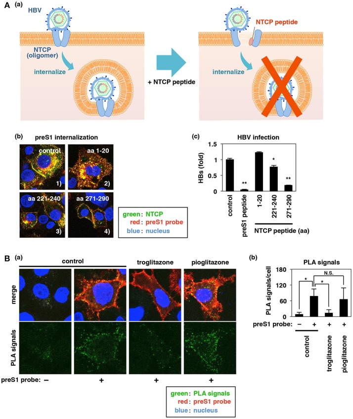
Figure 6.
Inhibition of NTCP oligomerization by peptides or troglitazone impedes HBV preS1-NTCP internalization. (Aa,b) Effect of NTCP peptides (aa 1–20, aa 221–240, and aa 271–290) on preS1 internalization as described in Figures 4Dc,d. Merged patterns of green (NTCP), red (preS1 probe), and blue (nucleus) signals are shown. Intracellular dotted yellow signals (the internalized preS1-NTCP) were clearly observed in the control and with aa 1–20, while those signals were decreased with aa 221–240 and even more strongly decreased with aa 271–290. (c) HBV infection of HepG2-hNTCP-C4 cells was examined as described in Figure 1B following treatment with or without the indicated peptides [preS1 or NTCP peptides (aa 1–20, aa 221–240, or aa 271–290)] using detection of HBs antigen at 12 days post-infection. (B) Effect of troglitazone on NTCP oligomerization was evaluated by PLA, as shown in Figure 5A. PLA signal was detected in HepG2 cells overexpressing both myc-NTCP and HA-NTCP following treatment with or without preS1 probe in the presence or absence of troglitazone or pioglitazone. (a) The lower panels show the PLA signal (green) only, and the upper panels are the merged images of green (PLA signal), red (preS1 probe), and blue (nucleus). (b) PLA signals in the experiment in (a) were quantified using Dynamic Cell Count (KEYENCE). Signal was significantly reduced by troglitazone, but not by pioglitazone. Data are shown as mean ± SD. Statistical significance was determined using a two-tailed non-paired Student's t-test (N.S.; not significant, *P < 0.05, **P < 0.01).

