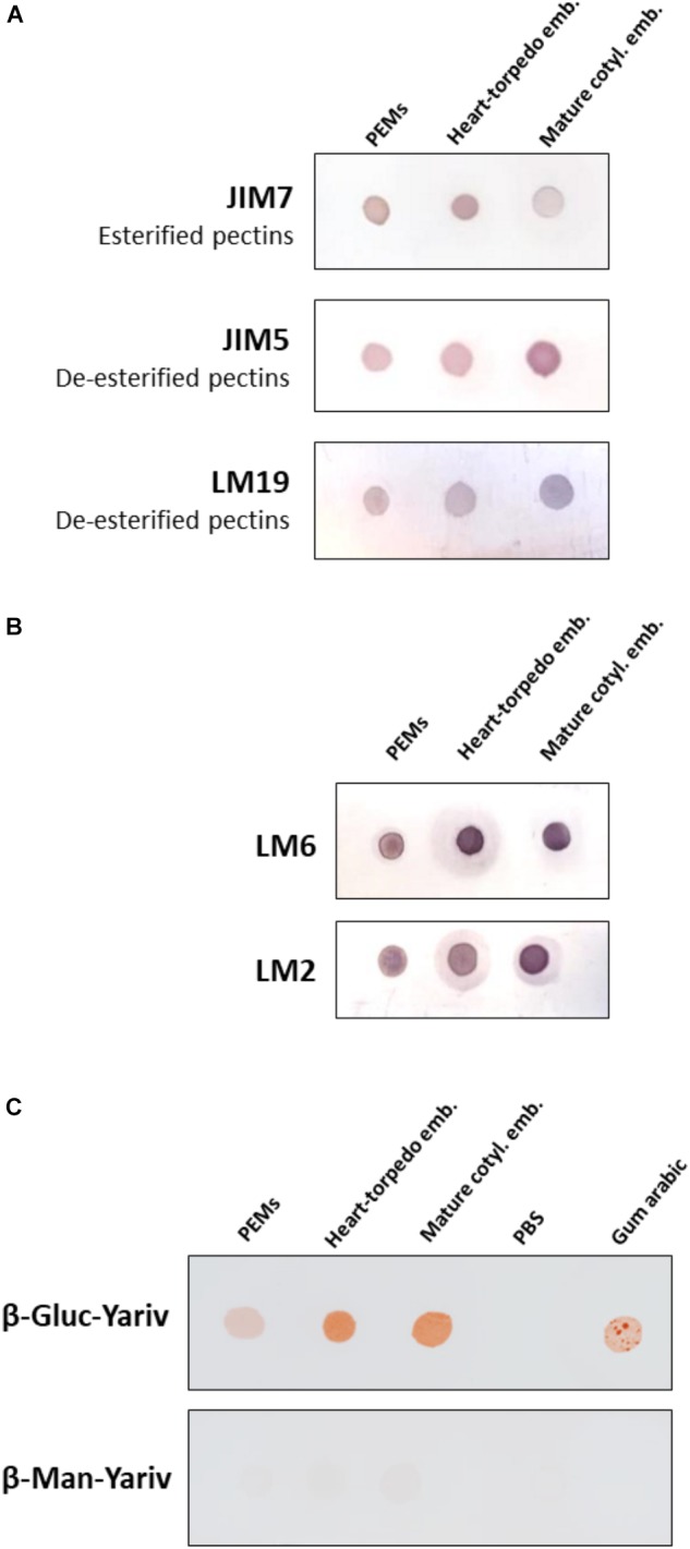
FIGURE 5.

Temporal profiles of esterified/de-esterified pectins and AGPs during somatic embryogenesis. Equal amounts of extracts were dotted for each developmental stage: proembryogenic masses (PEMs), heart-torpedo embryos, and cotyledonary embryos. (A) Immuno dot blot assays with JIM7 antibody (esterified pectins), JIM5 antibody (de-esterified pectins), and LM19 antibody (de-esterified pectins). (B) Immuno dot blot assays with LM6 and LM2 antibodies for specific AGP epitopes. (C) Dot blots stained by β-glucosyl-Yariv reagent, that binds to AGPs, and β-mannosyl-Yariv reagent, which does not bind AGPs; from left to right: PEMs, heart-torpedo embryos, cotyledonary embryos, PBS (negative control) and Gum arabic (positive control). Strip of β-Gluc-Yariv shows increasing color intensity (indicating increase of AGPs) during progression of somatic embryogenesis. Strip control stained with β-Man-Yariv does not provide color signal in any dot.
