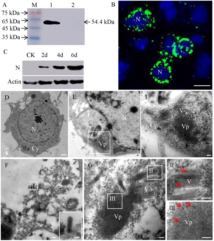
Figure 1.
Subcellular localization of protein N of RSMV in virus-infected VCMs. (A) Western blot analyses of N protein. Lane M, protein marker; lane 1, protein extracts from RSMV-infected leafhopper; lane 2, protein extracts from healthy leafhopper. (B) Immunofluorescence staining of N-FITC (green) and DAPI (4,6-diamidino-2-phenylindole) (blue) revealed punctate inclusions in cytoplasm of RSMV-infected VCMs at 48 h p.i. Bar, 100 μm. (C) Accumulation of N protein in 2, 4 and 6 days p.i. CK, mock-infected VCMs. (D) Electron micrograph of healthy cell of R. dorsalis. Bar, 2 μm. (E) Electron micrographs of RSMV-infected cell of R. dorsalis. Panel I was the enlarged image of the boxed areas in panel E. Viroplasm and virus particles of RSMV were distributed in cytoplasm. (F) Electron micrograph of virus particles distributed outside the cell. Insets show the image in the boxed area. (G) RSMV-infected VCMs were immunolabeled with N-specific IgG as the primary antibody, followed by treatment with 15-nm gold particle-conjugated goat antibodies against rabbit IgG as secondary antibodies. Panels II and III were the enlarged images of the boxed areas in panel G. Arrows indicate gold particles. Bars in panels E-G, I, II, and III, 100 nm.

