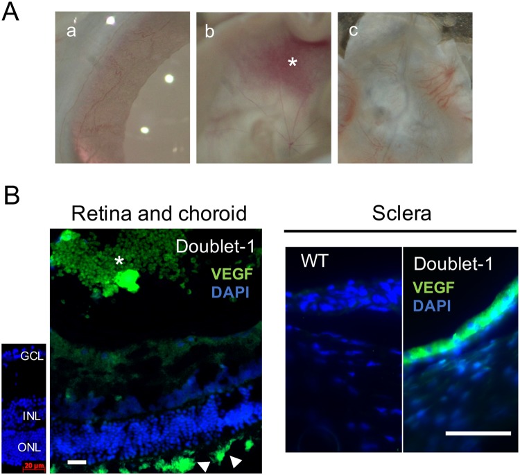
Figure 5.
(A) Representative photographies of iris, cornea, and sclera in Doublet-1 group. Neovascularizations of the retina, iris, and choroid were observed in 8 of 13 rats (62%) in the Doublet-1 treated eyes. (a) Iris, (b) Retina and epiretinal neovascular membrane (*), (c) Choroid and sclera (B) Representative photomicrographs of VEGF-immunopositive sclera and epiretinal membrane in the Doublet-1 treated eye. Intense VEGF immunopositive signaling was observed in the sections of sclera, choroidal vessels (arrowhead) and epiretinal neovascular membranes (*) Scale Bar: 20 μm.

