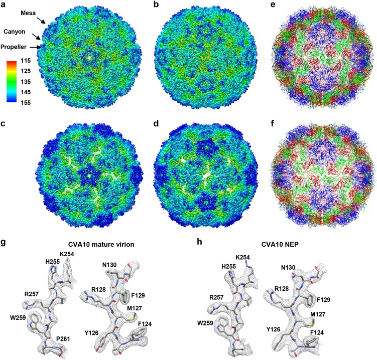
Fig. 1. Atomic-resolution cryo-EM structures of CV-A10 particles.
a, b Cryo-EM maps of CV-A10 mature virion viewed along the icosahedral five-fold (a) and two-fold (b) axis, respectively. The color bar labels the corresponding radius from the center of the sphere (unit in Å). The same color scheme was followed throughout. c, d Cryo-EM maps of CV-A10 NEP viewed along the icosahedral five-fold (c) and two-fold (d) axis, respectively. e, f Atomic models of CV-A10 mature virion (e) and NEP (f) viewed along the icosahedral two-fold axis, respectively. The models of capsid proteins VP1, VP2, VP3, and VP4 were colored in blue, green, red, and yellow, respectively. The same color scheme was followed throughout, unless otherwise indicated. g, h Atomic-resolution structural features of CV-A10 mature virion (g) and NEP (h), respectively, with the segmented density (mesh) in gray and the corresponding atomic model (sticks) in color. The well-resolved densities for almost all the side chains demonstrate the high resolution of the cryo-EM map

