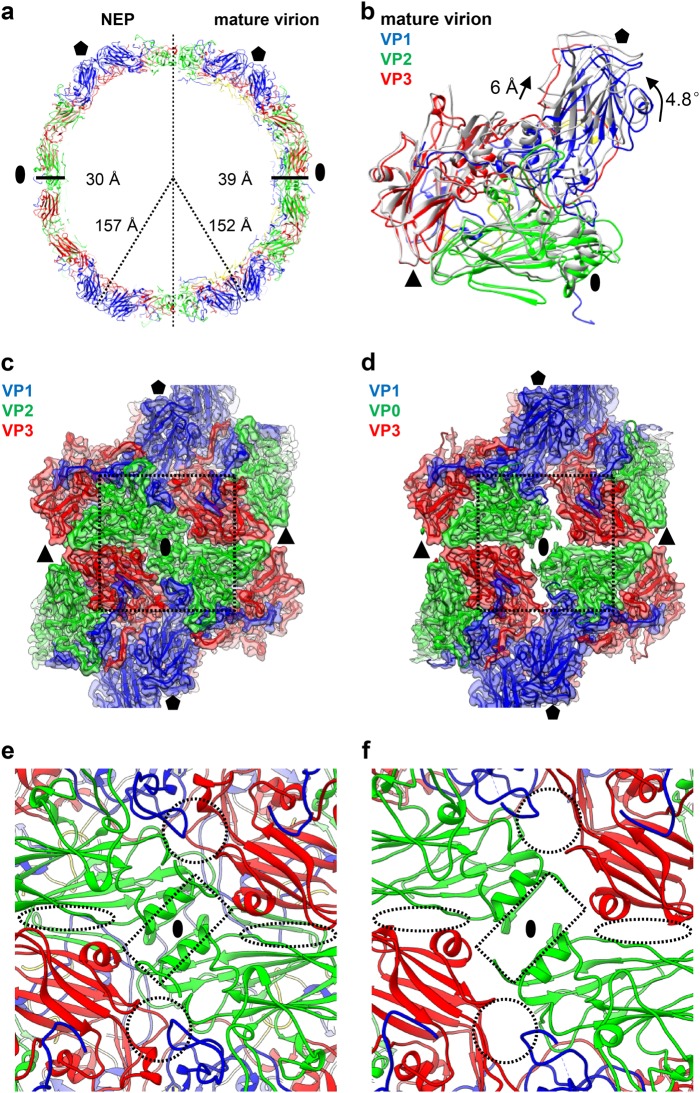
Fig. 2. Capsid structural comparison between CV-A10 mature virion and NEP.
a Two half sections of a 20 Å-thick central slab through the atomic models of CV-A10 NEP particle (left) and mature virion (right). Black oval and pentagon represent the two-fold and five-fold axes, respectively. The capsid radiuses and thickness for the two types of particles are also labeled. b One protomeric unit of CV-A10 NEP (in gray) was aligned with that of mature virion (in color). The rotation and translation from mature virion to NEP were also labeled. Black oval, triangle, and pentagon represent the two-fold, three-fold, and five-fold axes, respectively. c, d Structural configurations of four adjacent protomers around the two-fold axis for CV-A10 mature virion (c) and NEP (d), respectively. The major differences between them were indicated by dashed rectangle. e, f Zoom-in view of the icosahedral two-fold region of CV-A10 mature virion (e) and NEP (f). Dashed rectangle, circle, and oval indicate the locations of two-fold channel, a second channel nearby the quasi-three-fold axis, and another small ditch formed at the VP2/VP3 interface between adjacent protomers nearby the three-fold axis arising in the NEP, respectively

