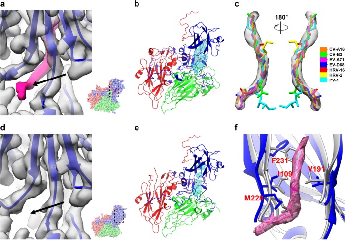
Fig. 4. VP1 pocket region and identification of pocket factor in the CV-A10 mature virion.
a Zoomed view of the VP1 pocket region in CV-A10 mature virion. The pocket location with respect to the complete CV-A10 protomer is illustrated in a small panel in the lower-right corner. The density corresponding to the pocket factor is highlighted in hot-pink. The entrance of the pocket is indicated by a black arrow. b The hydrophobic pocket (cyan mesh) in VP1 of CV-A10 mature virion is occupied by a natural lipid (hot-pink). c Fitting of putative pocket factor structures from seven known enterovirus structures (CV-A16: 5C4W, CV-B3: 1COV, EV-A71: 3VBF, EV-D68: 4WM8, HRV-16: 1AYN, HRV-2: 1FPN, and PV-1: 1VBD) into the corresponding density in the cryo-EM map of CV-A10 mature virion. d Zoom-in view of the VP1 pocket region in CV-A10 NEP. The same visualization style as in (a) was adapted. No density corresponding to the pocket factor can be observed. e The empty, collapsed pocket (cyan mesh) of CV-A10 NEP. f Comparison of the pocket region between CV-A10 mature virion (blue) and NEP (gray). Here the density and atomic model corresponding to the pocket factor are shown in hot pink

