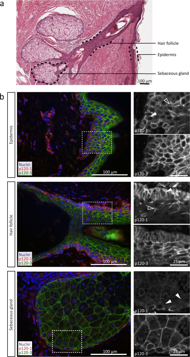
Figure 5.
p120 isoform localization in the skin. (a) Histological section of epidermis, containing a hair follicle and sebaceous glands. (b) p120-1 (6H11 mAb) and p120-3 expression (anti-p120-3 pAb) in the epidermis, the root sheath of a hair follicle and a sebaceous gland. Panels on the right represent a magnification of the area in the dashed boxes. Open and filled arrowheads indicate p120-1-positive cells in the basal layer (filled) and intermediate layers (open).

