Fig. 1.
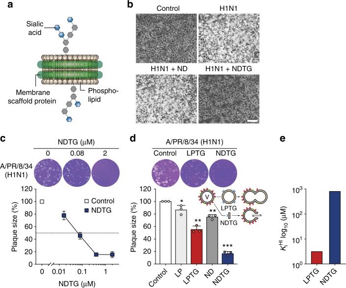
In vitro inhibitory activity of NDTG against H1N1 virus infection. a Cartoon representation of the total ganglioside-embedded nanodisc (NDTG). b Microscopy observations of virus-induced CPE reduction in MDCK cells by NDTG. MDCK cells were either uninfected (control) or challenged with A/PR/8/34 H1N1 virus (MOI = 0.1) at 25 °C for 24 h, with 0.5 μM ND or NDTG in the presence of TPCK-treated trypsin for activation of HA. CPE was observed at 24 h post-infection by inverted microscopy. Scale bar, 100 μm. c, d Evaluation of antiviral activity of NDTG against H1N1 virus by a plaque reduction assay. MDCK cells were infected with ~100 PFU of H1N1. One hour after infection, the viral inoculum was removed, and cells were overlaid with agarose containing the indicated concentrations of NDTG (c), or overlay supplemented with ND, NDTG, LP or LPTG at the same lipid concentration of 50 μM (d). Representative samples of viral plaques in each histogram are shown. Data are expressed as the mean ± standard deviation (SD) of triplicate samples. Error bars indicate SD, and asterisks indicate statistical significance determined by Student’s t-test (*P < 0.05; **P < 0.01; ***P < 0.001). e Hemagglutinin inhibition by NDTG or LPTG. The lowest lipid concentration of inhibitor that completely suppressed hemagglutination was defined as the hemagglutination inhibition constant KiHI

