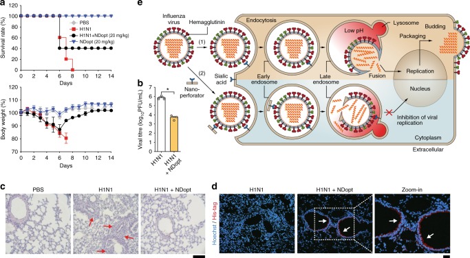
Fig. 5.
In vivo antiviral activity of NDopt. a Survival rates and body weight of the mice. NDopt (20 mg/kg) was intravenously administered to mice (n = 5/group) at 1 h after intranasal infection with mouse-lethal doses of H1N1 virus, and then once a day for 5 days. The survival rate (left) and body weight (right) of each group were monitored daily for 14 days. b–d Reduced viral replication and inflammation lesion by NDopt in the lungs of virus-infected mice. The lungs were obtained from mice administered intravenously with NDopt 5 days post-infection. b Reduced viral titres, as determined by a plaque assay, were observed in the lung administered with NDopt (n = 3). c Images of lung tissues stained by H&E for histopathologic examination. The representative micrographs from each group is shown at ×100 magnification. Red arrows indicate the presence of inflammatory cells in the tissue sections. Scale bars, 100 μm. d Representative fluorescence images of lung tissues of virus-infected mice treated with NDopt (×200 magnification). NDopt was detected using rabbit anti-His-tag antibody and Alexa Fluor 594-labelled anti-rabbit IgG antibody. White arrows indicate the presence of NDopt on the surface of the lung alveolar epithelial cells. Scale bars, 100 μm. e Proposed model for nano-perforator antiviral activity. Black arrows illustrate influenza virus infection pathway in the absence (1) or presence of nano-perforators (2). Data are expressed as the mean ± SD. Asterisks indicate statistical significance determined by Student’s t-test (*P < 0.05; **P < 0.01; ***P < 0.001) (a, b)

