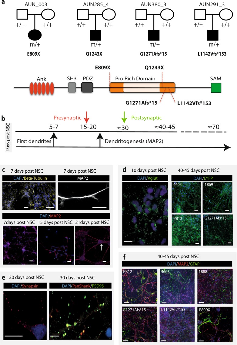
Figure 1.
Pedigrees of the families carrying de novo SHANK3 mutations and neuronal characterization. (a) Upper, the four patients probands carry de novo truncating mutations in SHANK3 gene (two « STOP » and two « frameshift » mutations leading to a premature STOP codon). (a) Lower, A schematic representation of the multidomain SHANK3 protein with the location of the four mutated aminoacids is provided. Conserved domains are indicated by filled rectangles. The mutations are located within the Proline-rich structure of SHANK3, between the PDZ and the SAM domains, and within exon 21 of SHANK3 gene. (b) Schematic representation of neuron maturation at different time periods. (c) Upper left, Immunofluorescence staining of human control iPSC cells-derived neurons by beta III tubulin at 7 days post NSC differentiation. (c) Upper right, labeling of a single dendrite using the MAP2 marker showing dendritic growth cones at early stages of culture. (c) Lower, Immunofluorescence staining of human iPSC cells-derived neurons by MAP2 at different intervals of time post NSC differentiation. The target shows neurite elongation and established connectivity between cell clusters. (d) VGlut1 marker at 10 days post NSC differentiation stained glutamatergic neurons. Labeling of control (1869, 4603, PB12) and ASD (G1271Afs*15) mature neurons at 40–45 days post NSC differentiation using the pAAV-CaMKIIa-hChR2-EYFP-WPRE lentivirus are also illustrated. (e) Immunofluorescence staining using presynaptic (synapsin) and postsynaptic (PSP95 and PanSHANK) markers. No staining was detectable before the indicated days post NSC differentiation (20 days for Synapsin, 10 days for VGlut1 and 30 days for PSD95). Data are from at least two control individuals (PB12 and 4603) (f) Immunofluorescence GFAP staining of cultures from iPSC-derived neurons showing the presence of astrocytes in 3 controls individuals (PB12, 4603, 1888) and 3 patients (G1271Afs*15, L1142Vfs*153, E809X). Scale bars: 10 μm (a-e); 100 μm (f).

