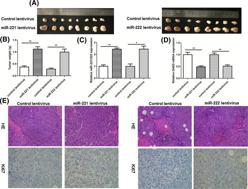
Figure 5. Effects of miR-221/222 on tumor growth in a breast cancer xenograft mouse model.
(A) MCF-7 cells were infected with a control lentivirus or lentiviruses to overexpress miR-221 or miR-222. After infection, MCF-7 cells (1 × 10⁷ cells per 0.1 ml) were subcutaneously implanted into 4-week-old nude mice (eight mice per group), and tumor growth was evaluated on day 25 after cell implantation. Representative images of the tumors from the implanted mice. (B) Quantitative analysis of the tumor weights. (C) Quantitative RT-PCR analysis of miR-221/222 levels in the tumors from implanted mice. (D) Quantitative RT-PCR analysis of GAS5 mRNA levels in the tumors from implanted mice. (E) Representative H&E-stained and Ki-67-stained sections of the tumors from implanted mice; *P<0.05; **P<0.01.

