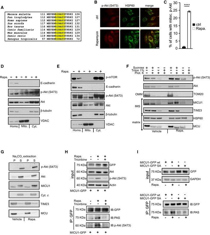
Figure 2. Mitochondrial Akt phosphorylates MICU1 at the Ser124 position.

- Sequence alignment of the MICU1 protein from nine vertebrate species. The Akt consensus phosphorylation motif, R‐X‐R‐X‐X‐S/T, is marked in yellow.
- HeLa cells treated with vehicle or 1 μM rapamycin for 4 h were stained for phosphorylated (S473) Akt (p‐Akt) or HSP60 (mitochondrial marker). Merged images are indicated (merge). Scale bar 10 μm.
- Analysis of the number of cells, expressed as a percentage, showing obvious mitochondrial staining of activated (S473 phosphorylated) Akt (p‐Akt) (n = 3 independent experiments; 350‐355 cells). Means ± SEM. ****P < 0.0001 (Student's t‐test).
- HEK293T cells treated with vehicle or 1 μM rapamycin (Rapa.) for 4 h were fractionated into cytosol (Cyt.) or mitochondrial (Mito.) extracts and analyzed by Western blotting. E‐cadherin: plasma membrane marker; β‐tubulin: cytosolic marker; VDAC: mitochondrial marker; Homo: cell homogenate.
- Mouse livers treated with vehicle or 1 μM rapamycin (Rapa.) for 16 h by an intraperitoneal injection were fractionated into cytosol (Cyt.) or mitochondrial (Mito.) extracts and analyzed by Western blotting. Phosphorylated (S2468) mTOR (p‐mTOR) was used to assess the rapamycin activity. E‐cadherin: plasma membrane marker; β‐tubulin: cytosolic marker; VDAC: mitochondrial marker; Homo: cell homogenate.
- Mitochondria isolated from HEK293T cells were subjected to the indicated treatments and analyzed by Western blotting against Akt, phosphorylated (S473) Akt (p‐Akt), and mitochondrial proteins with known localizations. Osmotic swelling through the removal of sucrose from the buffer was used to induce OMM rupture. OMM: outer mitochondrial membrane; IMS: intermembrane space.
- Western blot analysis of the supernatant (S) and insoluble pellet (P) fractions of vehicle‐ or rapamycin (Rapa., 1 μM for 4 h)‐treated HEK293T cell mitochondria following carbonate extraction at pH 11.5. Akt, phosphorylated (S473) Akt (p‐Akt), MICU1, the established integral membrane proteins TIM23 and MCU, and the soluble protein cytochrome c (Cyt. c) was analyzed.
- HEK293T cells were transfected with the GFP‐tagged wild‐type (WT) MICU1 and then treated with 1 μM rapamycin (Rapa.) alone for 4 h or in combination with the Akt inhibitor triciribine (10 μM). GFP‐MICU1 immunocomplexes were precipitated with a GFP antibody and analyzed with PAS (phospho‐Akt substrate) and phosphorylated (S473) Akt (p‐Akt) antibodies by Western blotting.
- HEK293T cells were transfected with either the GFP‐tagged wild‐type (WT) MICU1 or MICU1 S124A‐GFP mutant. MICU1‐GFP immunocomplexes were precipitated with a GFP antibody and analyzed with PAS (phospho‐Akt substrate) antibody by Western blotting.
Source data are available online for this figure.
