-
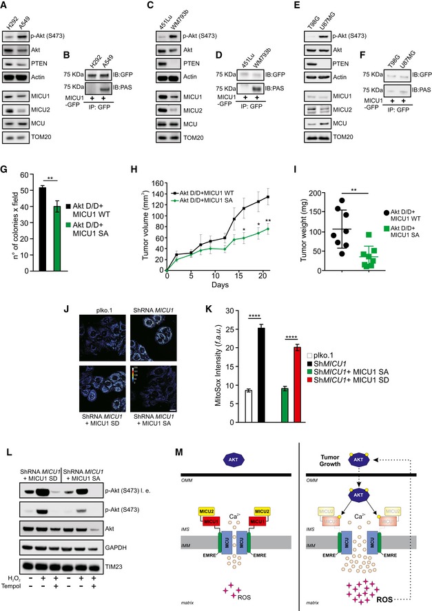
A
Western blot analysis of MICU1 and MICU2 levels in lung cancer cells with low or high Akt activity.
-
B
Lung cancer cells were transfected with the GFP‐tagged WT MICU1. MICU1‐GFP immunocomplexes were precipitated with a GFP antibody and analyzed with a PAS (phospho‐Akt substrate) antibody by Western blotting.
-
C
Western blot analysis of MICU1 and MICU2 levels in melanoma cells with low or high Akt activity.
-
D
Melanoma cells were transfected with the GFP‐tagged WT MICU1. MICU1‐GFP immunocomplexes were precipitated with a GFP antibody and analyzed with PAS (phospho‐Akt substrate) antibody by Western blotting.
-
E
Western blot analysis of MICU1 and MICU2 levels in glioblastoma cells with low or high Akt activity.
-
F
Glioblastoma cells were transfected with the GFP‐tagged WT MICU1. MICU1‐GFP immunocomplexes were precipitated with GFP antibody and analyzed with PAS (phospho‐Akt substrate) antibody by Western blotting.
-
G–I
Number of soft agar colonies (n = 3 independent experiments) (G), xenograft tumor volumes (H), and tumor weights at day 21 (n = 8 mice for each group) (I) formed by Rat2 cells stably expressing the indicated constructs.
-
J, K
Representative images (J) and analysis (K) of MitoSOX‐based ROS measurements in ShRNA MICU1 HeLa stable cells stably expressing either MICU1 S124D (SD) or MICU1 S124A (SA) (n = 3 independent experiments). Scale bar 10 μm. f.a.u.: fluorescence arbitrary unit.
-
L
ShRNA MICU1 HeLa stable cells stably expressing either MICU1 S124D (SD) or MICU1 S124A (SA) were treated with 500 μM H2O2 alone or in combination with the ROS scavenger Tempol and analyzed by Western blotting as indicated. l. e.: long exposure.
-
M
Speculative model of the mitochondrial Akt‐MICU1 axis in the regulation of tumor growth.
Data information: (G, I) Means ± SEM. **
‐test); (H) Means ± SEM. *
‐tests); (K) Means ± SEM. ****
< 0.0001 (one‐way ANOVA).