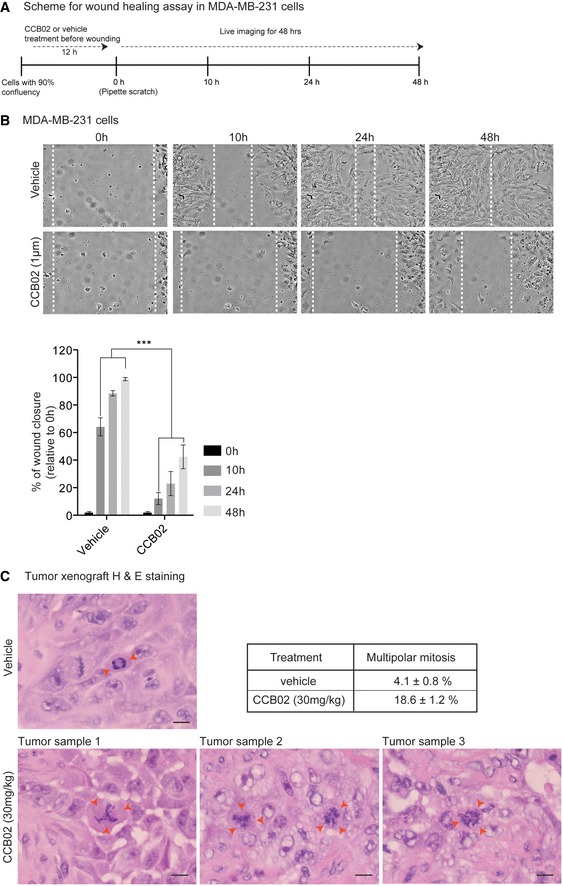
Figure EV5. Related to Fig 8: CCB02 prevents MDA‐MB‐231 cell migration and induces multipolar mitosis in mouse xenografts.

- Experimental scheme of wound‐healing assay using MDA‐MB‐231 cells.
- Snapshot of live cell images shows wound closure at various time points (0, 10, 24, and 48 h). Relative to vehicle treatment, CCB02 delays wound closure. Dashed lines mark cell‐free empty space. Scale bar, 100 μm. Bar diagrams at right quantify the percentage of relative wound closure. (N) = 3. Error bars, mean ± SEM. P‐values were obtained using two‐way ANOVA. ***P < 0.0001.
- Image shows H&E immunohistochemistry staining of representative xenograft tumor samples. In contrast to vehicle control xenograft tumors that show bipolar spindles (red arrows, top panel), CCB02 xenograft‐treated tumors show increased frequencies of multipolar mitotic cells, indicating mitotic catastrophe (red arrows, bottom panel). Scale bar, 10 μm. Table shows the percentage of multipolar mitotic cells. Data are represented as mean ± SEM. At least 350 mitotic cells were scored for vehicle and CCB02 treatment from independent xenograft tumors.
