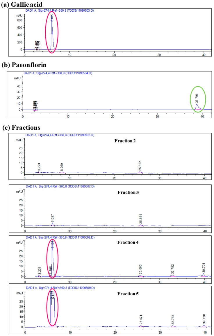
Figure 2.
HPLC profiles of bioactive markers (a) gallic acid; (b) paeoniflorin and (c) fractions 2, 3, 4 and 5 of Cortex Moutan HSCCC extract. Detection was performed at UV 274 nm. Representative retention time peaks of the bioactive markers are highlighted in circle. Fractions 4 and 5 showed retention time peak similar to that of gallic acid (highlighted in circle).

