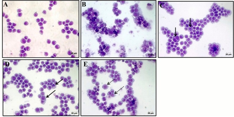
Figure 4.
Effect of 16 on cell morphology for HL-60 human leukemia cells. The cells were stained with hematoxylin-eosin and analyzed by optical microscopy after 24 h incubation at concentrations of 0.32 (C); 0.64 (D); and 1.28 μg/mL (E). Negative control (A) was treated with the vehicle (0.1% DMSO) used for diluting the test substance. Doxorubicin (0.3 μg/mL) was used as the positive control (B). Continuous arrows show nuclear fragmentation and non-continuous arrows show accumulation of metaphases cells.

