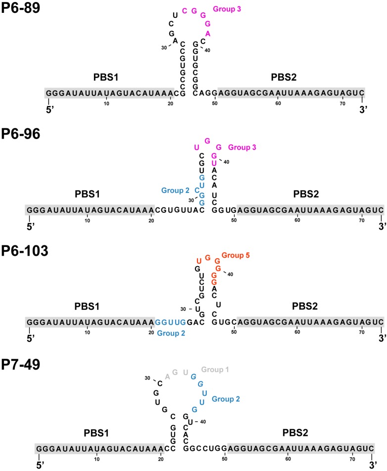
Figure 3.
Proposed secondary structure of P6-86, P6-96, P6-103 and P7-49 as determined by TurboFold software. Theoretical nucleotide motifs involved in the interaction with the CRE are colored according to the group they belong to, as indicated in Figure 1B. The constant and common sequences for all the aptamers tested, PBS1 and PBS2, are highlighted in grey. PBS, primer binding site.

