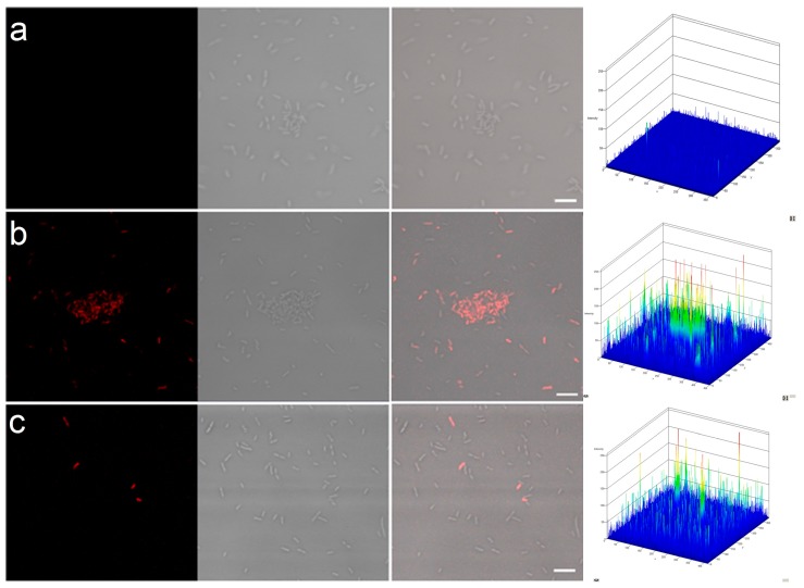
Figure 6.
Permeability of cell membranes of P. aeruginosa after treatment with PPI dendrimers.
(a) control; (b) PPI 1 µM with AMX 1 µg/mL; (c) PPI 100%malG3 5µM with AMX 1 µg/mL. For all photomicrographs the left row shows red fluorescence (propidium iodide, PI, staining) and the right shows Nomarski differential interference contrast.

