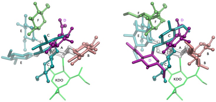
Figure 8.
Overview on conformations found for the highly branched glucose-rich inner core of lipooligosaccharide of M. catarrhalis. (Left) Comparison of the energy minimum conformation of lgt1/4Δ OS (stick) and the “(1-4)anti-ψ(1-6)gg” conformation of lgt2Δ OS (cpk); (Right) Comparison of the “(1-4)anti-ψ(1-6)gg” conformation (cpk) of lgt2Δ OS and a conformation that has the (1-4)-linkage in minimum A and the (1-3)-linkage in minimum B. The terminal GlcNAc residue (D) is shown in purple.

