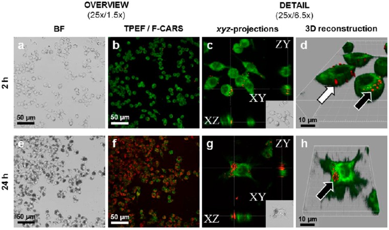
Figure 3.
Interactions between paliperidone palmitate (PP) nanocrystals and RAW 264.7 macrophages imaged by CARS after 2 (a–d) and 24 h (e–h). Imaged is performed using CARS signal at 2860 cm−1 and fluorescently dyed cell membranes are imaged using TPEF. (a,e) low and high magnification brightfield imaging; (b,f) forward-CARS (red)/TPEF (green) merged micrographs of stained/fixed cells; In (c,g) intracellular PP nanocrystals are seen in orthogonal projections of z-stacked F-CARS/TPEF overlays; (d,h) show 3D-reconstructions of the z-stacked F-CARS/TPEF overlays. White arrows indicate PP-NC adsorbed onto cell surface and black arrows phagocytosed PP-NCs. (From [21], reprinted from Elsevier with permission).

