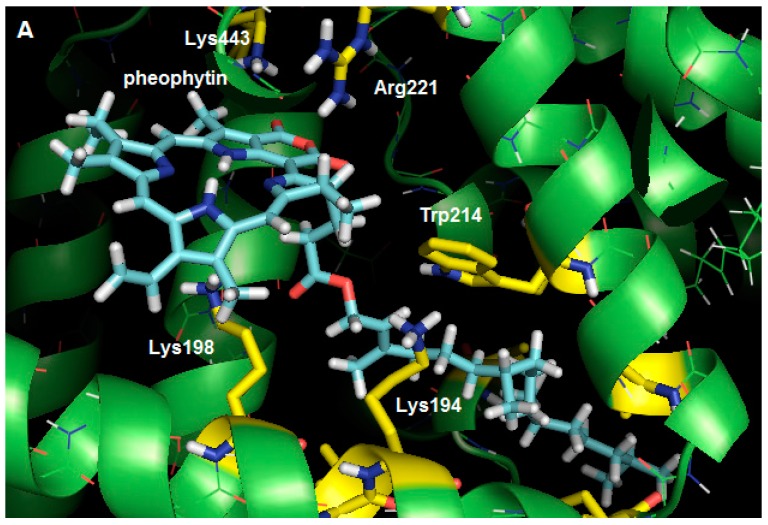
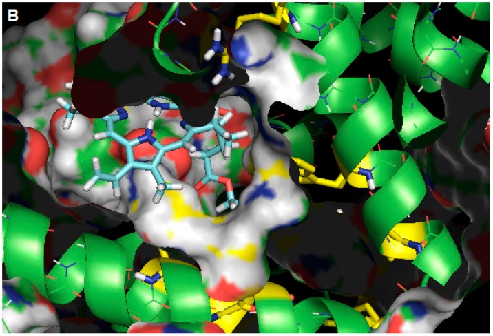
Figure 7.
(A) Best score pose for pheophytin in HSA, obtained by molecular docking (ChemPLP function). Carbon: cyan (pheophytin), green (HSA), yellow (selected residues); hydrogen: white; oxygen: red; and nitrogen: blue; (B) Representation of the molecular surface of HSA, where it can be seen that the apolar side chain of pheophytin is completely surrounded by the hydrophobic gorge (figures generated with the PyMOL software).


