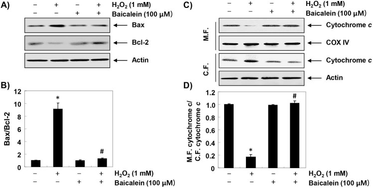
Figure 6.
Effects of baicalein on H2O2-induced changes of Bax and Bcl-2 expression in RT4-D6P2T cells. (A and C) Cells were treated with 100 µM baicalein for 1 h, and then stimulated with or without 1 mM H2O2 for 24 h. The cellular proteins were separated by SDS-polyacrylamide gel electrophoresis, and then transferred to membranes. The membranes were probed with the indicated antibodies. Proteins were visualized using an ECL detection system. Actin was used as an internal control. (C) The mitochondrial and cytosolic proteins isolated from cells cultured under the same conditions were separated by SDS polyacrylamide gel electrophoresis, and transferred to the membranes. The membranes were probed with anti-cytochrome c antibody. The proteins were visualized using an ECL detection system. Equal protein loading was confirmed by the analysis of COX VI and actin in each protein extract. (B and D) Bands were quantified using ImageJ and normalized to actin or COX IV, and the ratio was determined. Data are expressed as mean ± SD. All experiments were repeated three times (#p<0.05 in comparison to the control group; *p<0.05 compared with the H2O2 group).

