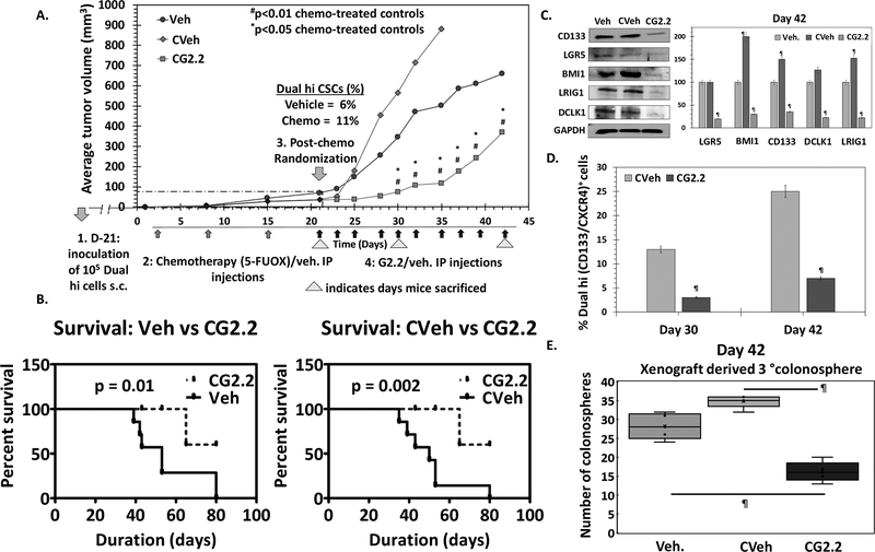
Figure 3: G2.2 inhibits CSCs in chemotherapy-enriched xenograft model.
(A) 5-flurouracil (25mg/kg) and oxaliplatin (2mg/kg) (FUOX) treatment (weekly × 3 weeks i.p.) of HT-29 Dual hi induced xenograft caused a modest reduction in tumor volume but significant enrichment of Dual hi CSCs. Treatment with G2.2 (200mg/kg 3 times/week × 10 injections i.p.) introduced at day 21 post-FUOX treatment-initiation produced significant reduction in tumor volume compared to Veh and CVeh. The arrows under the growth curves represent G2.2 treatment. (B) Bar graph representation of the flow cytometric analyses to determine proportion of Dual hi cells show significant reduction of CSCs by CG2.2 compared to CVeh. (C) FUOX treated xenograft-derived cells (CVeh) showed significant increase in CSC self-renewal (4° sphere formation) which was promptly inhibited by CG2.2 compared to both Veh. and CVeh treatments. (D) Western blot analyses and the corresponding bar graphs showing relative densitometric values normalized to GAPDH demonstrate inhibition of CSC (LGR5, CD133, LRIG1, DCLK1) and self renewal (BMI1) markers compared to both Veh and CVeh controls. Of note, CVeh xenograft showed incresed expression of CSC/self-renewal markers compared to Veh controls. (E) CG2.2 treated xenograft-derived cells show significant inhibition of CSC self-renewal (3° sphere formation) compared to both Cveh and Veh controls. Error bars represent ±1 SEM. #p-value < 0.01 compared to CVeh treated control and *p value <0.05 compared to Veh treated control. ¶p-value < 0.005 compared to Veh treated control.

