Fig. 4.
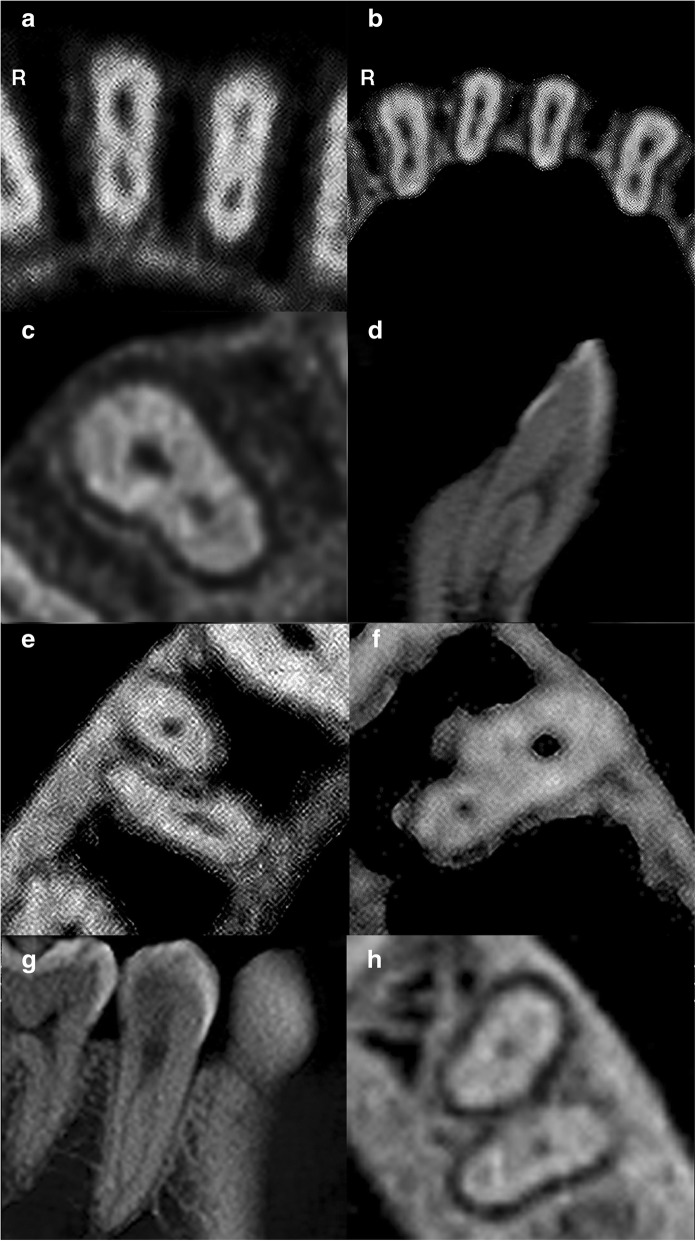
CBCT images of mandibular teeth. R – Right. a Axial slice of mandibular central incisors displaying two canals. b Axial slice of mandibular lateral incisors displaying two canals and canines displaying one canal. c Axial slice of a right mandibular canine displaying two canals. d Sagittal slice of a two-rooted left mandibular canine displaying two canals. e Axial slice of a two-rooted right mandibular first premolar displaying one canal in each root. f Axial slice of a left mandibular first premolar displaying two canals. g Sagittal slice of a single-rooted right mandibular second premolar displaying two canals. h Axial slice of a two-rooted left mandibular second premolar displaying one canal in each root

