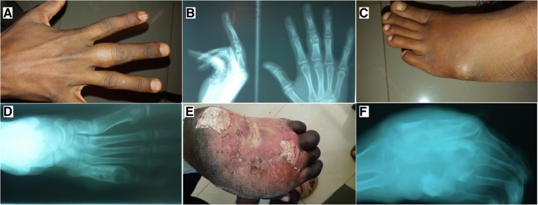
Fig. 2.
Pre-amputation images of the finger, toe, and hand with X-ray showing mycetoma. a Right middle finger eumycetoma (proximal phalanx). b PA and lateral X-ray view of the same patients showing cavity and evidence of mycetoma (this patient treated by finger Ray’s amputation). c Left toe mycetoma with sinus cavity. d X-ray was showing cavity and mycetoma (this patient treated by toe Ray’s amputation). e Hand showing mycetoma deformity, discoloration, and loss of function. f X-ray of the same patient was showing a deformity, cavitation, and mycetoma (this patient treated by below-elbow amputation)

