
Fig. 2.
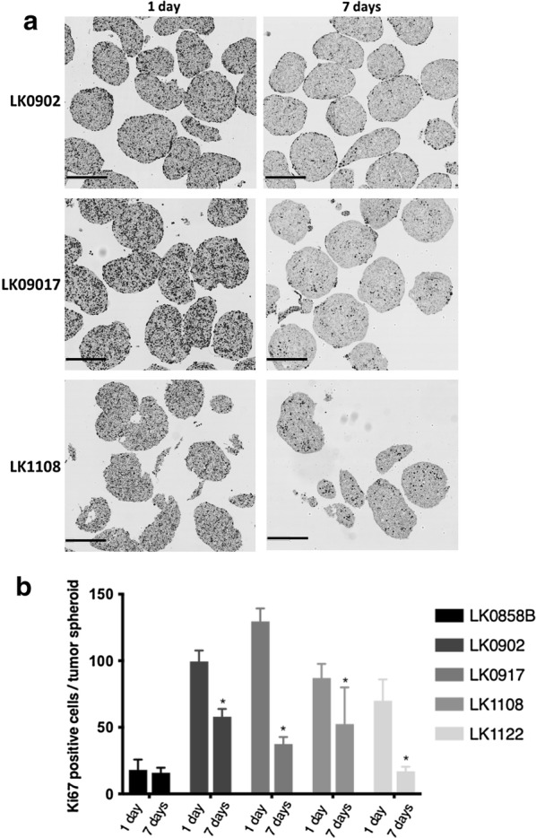
Histological evaluation of HNSCC-derived tumor spheroids. a Immunohistochemical staining of HNSCC tumor spheroids with the proliferation marker Ki67 cultured for 1 and 7 days; scale bar = 300 µm. b Quantification of Ki67-positive cells in 1 day and 7 days old tumor spheroids; the data are depicted as mean of ± SD, n = 10. *p < 0.05 according to Student’s test