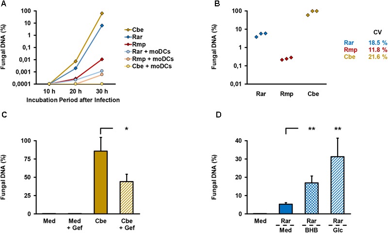
FIGURE 2.

Validation of endothelial compartment invasion and its reproducibility. (A) The bilayer model was assembled as described in the Section “Materials and Methods”. 2.5 × 10⁵ spores of different Mucorales species ± 2.5 × 10⁵ moDCs were added to the upper compartment. Mucorales DNA in the lower compartment was quantified using an 18S-based assay. The diagram shows the percentage of detected DNA in the lower compartment depending on incubation time after infection of the upper compartment. The percent scale refers to a positive control (=100%) incubated for 30 h after addition of the same number of fungal spores directly to the lower compartment. Mean values based on technical duplicates from one representative run are shown. Rar, R. arrhizus; Rmp, R. pusillus; Cbe, C. bertholletiae. (B) To document the reproducibility of endothelial compartment invasion, the model was assembled and infected with spores of each pathogen in three independent experiments. Mucorales DNA was quantified after 30 h and compared with positive controls as described in (A). Mean values of technical duplicates in each run are shown and inter-assay CVs are provided. Intra-assay CVs were consistently < 10%. (C) Endothelial compartment invasion by C. bertholletiae was assessed in the presence or absence of 50 μg/ml gefitinib (Gef) in the lower chamber. Uninfected inserts (medium control, Med) with and without gefitinib served as negative controls. Mucorales DNA was quantified after 30 h and compared with positive controls as described in (A). Three independent experiments were performed. Mean values and standard deviations are shown. (D) Prior to infection with R. arrhizus (Rar), Transwell® inserts assembled as described in the Section “Materials and Methods” were transferred to new wells containing either regular HPAEC medium (Med, glucose concentration: 1 mg/ml) or HPAEC medium supplemented with 8 mg/ml glucose (Glc) or 1 mg/ml beta-hydroxybutyrate (BHB). Mucorales DNA in the lower compartment was quantified 30 h post-infection and normalized to a positive control (=100%) as described above. Mean values from four independent experiments and standard deviations are shown. The two-sided Student’s t-test was used for significance testing in (C,D). ∗p < 0.05, ∗∗p < 0.01.
