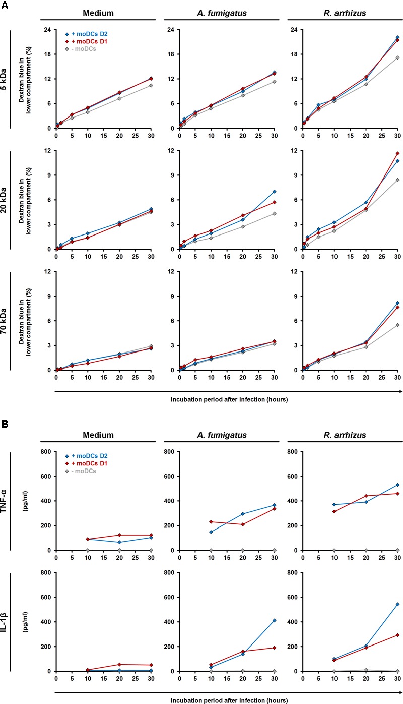
FIGURE 7.

Influence of dendritic cells on alveolar barrier dysfunction caused by A. fumigatus and R. arrhizus. (A) Dextran blue assays were performed as described in the Section “Materials and Methods” and figure legend 6. 2.5 × 10⁵ moDCs from two healthy donors (red and blue diamonds) or plain medium (gray diamonds) were added to the dextran blue solution. Medium (left column), A. fumigatus conidia (central column), and R. arrhizus spores (right column) were used for infection. (B) In parallel, concentrations of TNF-α and IL-1β were determined in inserts without dextran blue 10, 20, and 30 h after infection. (A,B) Dextran blue assays and ELISA were performed in technical duplicates, and mean values are shown in the figure (CVs consistently < 20%).
