Figure 1.

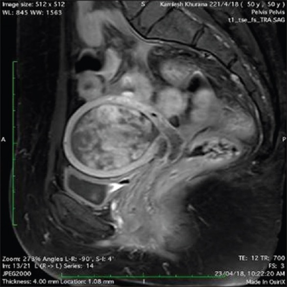
T1-weighted sagittal postcontrast image showing uterine cavity filled with heterogeneous mass lesion invading half of the myometrium. Cervix appearing normal

T1-weighted sagittal postcontrast image showing uterine cavity filled with heterogeneous mass lesion invading half of the myometrium. Cervix appearing normal