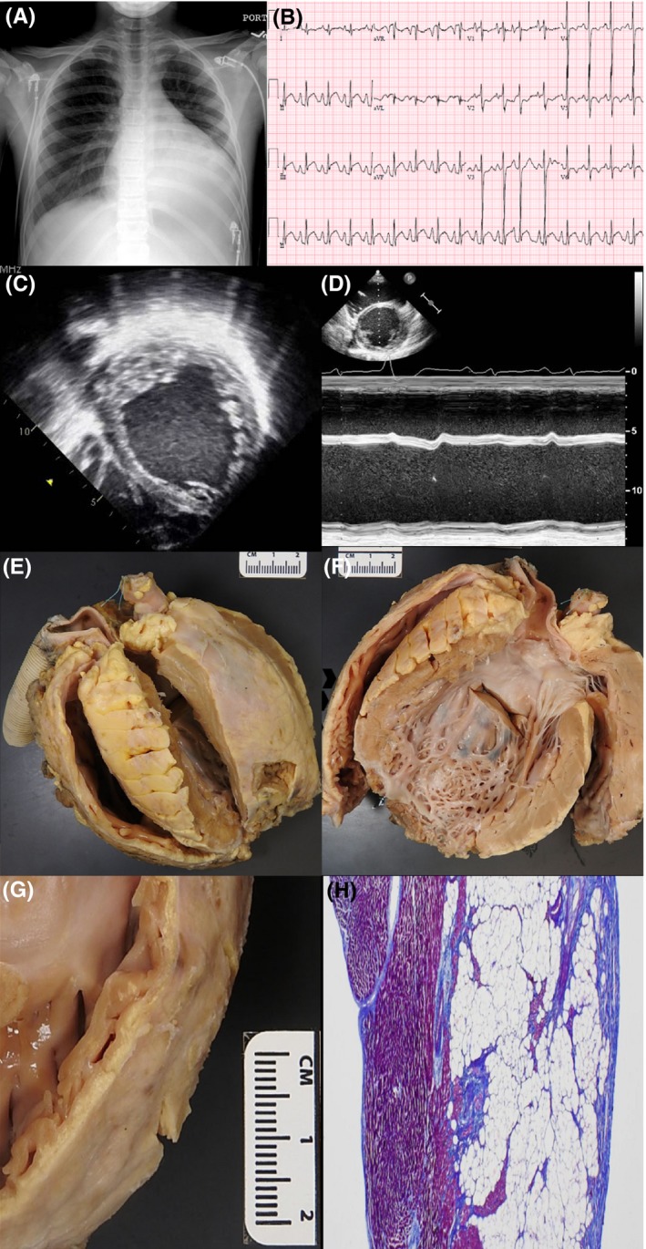
Figure 1.

A, Chest X‐ray demonstrates cardiomegaly, pulmonary congestion, and left pleural effusion. B, ECG demonstrates sinus tachycardia with premature atrial complexes, biatrial enlargement, ST elevation in inferior leads, and T‐wave inversion in lateral leads. C and D, Echocardiogram demonstrates severely dilated left and right ventricles with prominent left ventricular trabeculations. E, Gross anatomy of explanted heart demonstrates dilated thin‐walled RV and dilated globular LV. F, LV chamber demonstrates severe dilation and a hypertrebeculated LV apex and free wall. Left ventricular assist device insertion site excision is seen at the apex. G and H, High‐resolution gross image and histologic view of the RV free wall shows fibro‐fatty infiltration of the myocardium with sparing of the subendocardium
