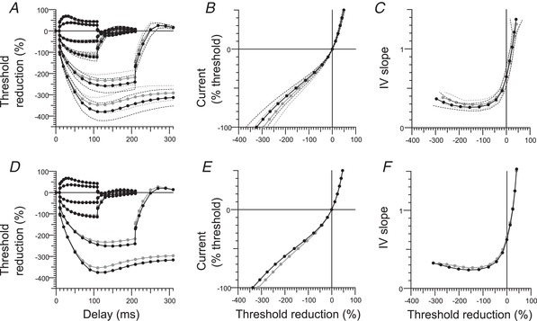
Figure 4. Evidence for greater I h in median nerve with the best‐fit from the mathematical model.

Extended excitability data for motor axons in the median nerve. A, B and C, upper row: group data (mean and SD); normal controls (n = 38; black filled circles) and RLS patients (n = 34; grey filled circles) and best‐fit from the Bostock motor axon model (D, E and F; lower row: black circles indicate the model in control subjects, and the modelled changes in RLS patients are shown as grey circles). A and D, TE for conditioning levels of +20%, –20%, +40%, –40%, –70% and –100% of control threshold. B and E, I–V for 200‐ms conditioning stimuli. C and F, I–V slope (threshold conductance): resting I–V slope (r): calculated from currents between –10% and +10%, shown at x = 0; minimal I–V slope.
