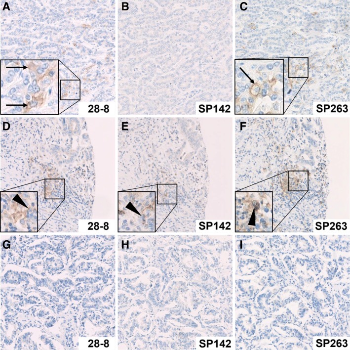
Fig. 1.
Examples of PD-L1 staining in a representative intrahepatic cholangiocarcinoma. The different staining characteristics of PD-L1 clones 28–8, SP142 and SP263 are displayed. In the first row, tumor cells show membranous positivity of few tumor cells with antibody clones 28–8 (a) and SP263 (c), but not with clone SP142 (b). In the second row, stromal inflammatory cells show membranous PD-L1 expression with all three antibody clones (d-f), while tumor cells are negative. In the third row, all samples are negative, both in tumor and stromal cells, and with all three PD-L1 antibodies (g-i). Original magnification: 200x, PD-L1 positive tumor cells are highlighted by black arrows, PD-L1 positive stromal cells are highlighted by black triangles

