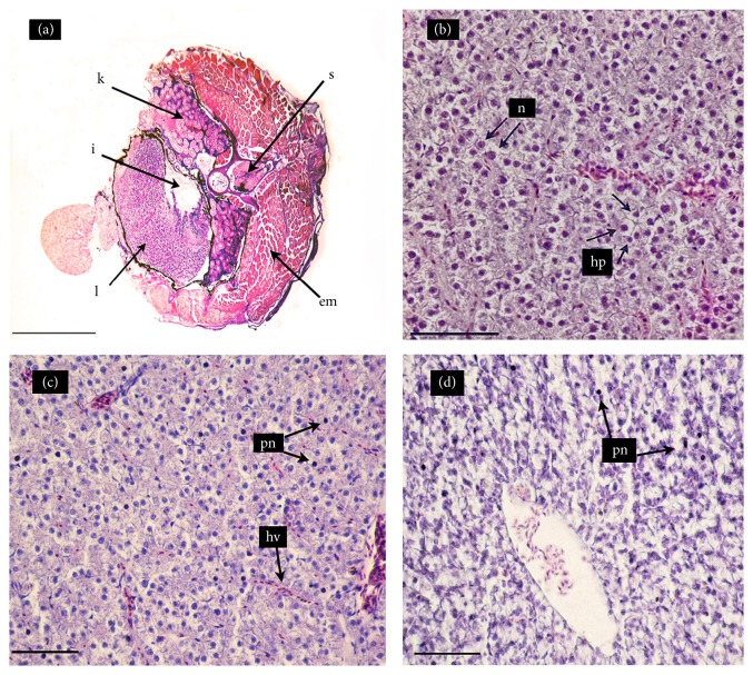
Figure 2.
Hematoxylin-eosin stained cross-sections of the mid-section of Cnesterodon decemmaculatus exposed to different treatments for 96 h. Treatments: NC (negative control, MHW); RR (surface water of the Reconquista River); RR+Cd (surface water of the Reconquista River with 2 mg Cd/L). (a) Fish exposed to NC (5X): liver (L); intestine (I); kidney (K); epiaxial muscle (EM); and spine (S). (b) Liver of fish exposed to NC (40X): normal hepatocytes (hp) with one nucleus (n) each. (c) Liver of fish exposed to RR (40X): hepatic vein (hv) and hepatocytes with pyknotic nuclei (pn) in the parenchyma and thin sinusoids in the parenchyma. (d) Liver of fish exposed to RR+Cd (40X): marked increase in the number of hepatocytes with pyknotic nuclei (pn).

