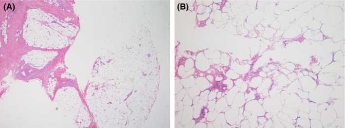
Figure 2.

The histopathology from left thigh showed (A) lobular panniculitis with sparse cells infiltration (Hematoxylin‐eosin staining, ×4) (B) lymphocytic and histiocytic infiltrations with some foci of lipomembranous change (Hematoxylin‐eosin staining, ×40) 541 × 203 mm (150 × 150 DPI)
