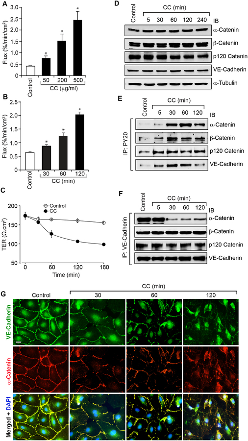
Figure 1. Cholesterol crystals disrupt endothelial AJ integrity and its barrier function.
A. Quiescent HAEC monolayer was treated with and without the indicated concentrations of cholesterol crystals for 2 hrs and flux was measured. B & C. Quiescent HAEC monolayer was treated with and without cholesterol crystals (200 mg/ml) for the indicated time periods and flux (B) and TER (C) were measured. D-F. Quiescent HAEC were treated with and without cholesterol crystals (200 mg/ml) for the indicated time periods, cell extracts were prepared and equal amounts of proteins from control and each treatment were either analyzed by Western blotting (IB) for the steady state levels of the indicated proteins (D) or immunoprecipitated (IP) with PY20 or VE-cadherin antibodies and the immunocomplexes were analyzed by immunoblotting (IB) for the indicated proteins (E & F) using their specific antibodies. G. Quiescent HAEC were treated with and without cholesterol crystals (200 μg/ml) for the indicated time periods and examined for AJ integrity by double immunofluorescence staining for VE-cadherin and α-catenin. Nucleus was stained with DAPI. The bar graphs represent quantitative analysis of three experiments. The values are expressed as Means ± SD. *, p < 0.05 vs control.

