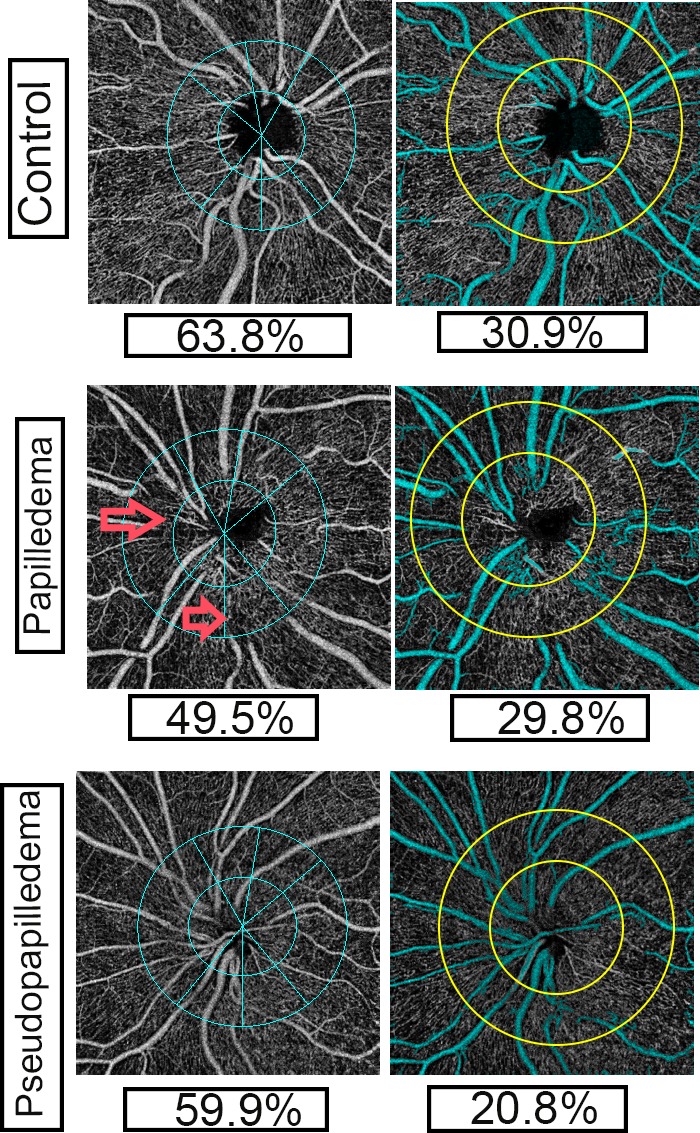
Figure 2.

OCT-A of peripapillary total vasculature (left column) and capillaries (right column) of inner retinal images in control, papilledema, and pseudopapilledema eyes. Left column: Commercial OCT-A (Optovue) sectors of peripapillary vasculature with peripapillary total vasculature density shown. Right column: OCT-A images with two concentric circles with 3.45- and 1.95-mm-diameter with customized software; major vessel (in cyan) removed using customized software. Peripapillary capillary densities were also shown. Capillary density in pseudopapilledema eyes is lower than papilledema and control eyes. Large vessel distortions and obscurations are seen in OCT-A images of papilledema (red arrows) compared with pseudopapilledema eyes.
