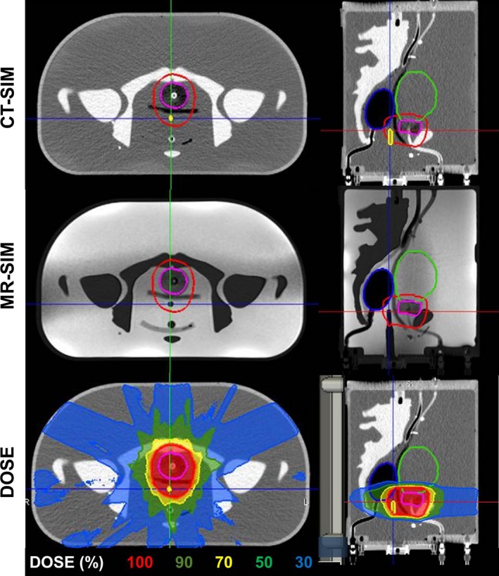
Figure 8.

Treatment plan: axial and sagittal views of the CT‐SIM, 0.35 T MR‐SIM, and dose for an 11‐field IMRT 6XFFF MR‐Linac treatment plan at the same slice in the phantom. Contours of the bladder (green), rectum (blue), gross tumor volume (pink), planned target volume (red), and ion chamber (yellow) are shown. The chamber is not delineated in the MR‐SIM to highlight its visibility for localization.
